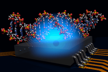
Quantum MRI Machine Allows Single Molecule Microscopy
By Jof Enriquez,
Follow me on Twitter @jofenriq

Australian scientists have successfully used quantum bits (qubits) to act as a miniaturized magnetic resonance imaging (MRI) sensor to image the atomic structure of a single molecule – a formidable challenge in the physical biosciences. Such a quantum molecular microscope could help researchers better understand biological processes and lead to the discovery of new drugs.
Usually associated with quantum computers, qubits – a basic unit of information capable of existing in two states, 0 or 1, simultaneously – can also be utilized as highly sensitive quantum sensors to image a biomolecule.
"Determining the structure of bio-molecules such as proteins can often be a barrier to the development of novel drugs," says Prof. Lloyd Hollenberg, Thomas Baker Chair in Physical Biosciences at the University of Melbourne, in a news release. "By using quantum sensing to image individual atoms in a bio-molecule, we hope to overcome several issues in conventional biomolecule imaging."
Existing state-of-the-art techniques to image molecules – such as X-ray crystallography, nuclear magnetic resonance (NMR) spectroscopy, or free electron laser femtosecond serial crystallography – depend on large numbers of the target molecule to be crystallized and subjected to X-ray diffraction to determine the molecule's average structure.
However, not all biomolecules are amenable for crystallization, particularly proteins associated with cell membranes, which are critical in the development of new drugs. Those that survive the process tend to lose vital information about their full molecular structure.
Therefore, researchers at the University of Melbourne and at the ARC Centre of Excellence for Quantum Computation and Communication Technology (CQC2T), developed a new system that relies instead on "a quantum spin probe acting simultaneously as the magnetic resonance sensor and source of magnetic field gradient" which encodes signals corresponding to specific regions of the molecule’s nuclear spin density to produce a 3D image of the target molecule rapamycin.
"In a conventional MRI machine large magnets set up a field gradient in all three directions to create 3D images; in our system we use the natural magnetic properties of a single atomic qubit," explains University of Melbourne PhD researcher Viktor Perunicic, who was the lead author on the paper published in Nature Communications.
"The system would be fabricated on-chip, and by carefully controlling the quantum state of the qubit probe as it interacts with the atoms in the target molecule, we can extract information about the positions of atoms by periodically measuring the qubit probe and thus create an image of the molecule's structure," adds Perunicic.
The scientists say the molecules hydrogen and carbon substructure were successfully imaged at the angstrom level, and that their quantum MRI machine can scale up to image biomolecules considerably larger than rapamycin. They used diamond-based qubits in their experiment, but silicon-based qubits in a future atomic-scale MRI could offer even higher resolution images of a single molecule, which bodes well for medicine.
"The construction of such a quantum MRI machine for single molecule microscopy could revolutionise how we view biological processes at the molecular level, and could lead to the development of new biotechnology and a range of clinical applications," says Prof. Hollenberg.
