How To Prepare Your Specimen For Immunofluorescence Microscopy
Source: Danaher Life Sciences
By Florian Hoff, Leica Microsystems

This content is brought to you by Leica Microsystems, a Danaher Operating Company.
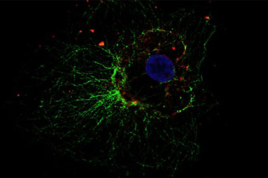
Immunofluorescence (IF) has become indispensable for many research groups who have at least access to a simple fluorescence microscope. It is a powerful method for visualizing intracellular processes, conditions and structures. The centerpiece of an Immunofluorescence (IF) experiment is a combination of two different components:
- First, specific antibodies, which are used to form an immune complex to mark the desired molecules – in most cases proteins – in the cell.
- Second, fluorochromes, which are coupled to the immune complexes and therefore visualize the target structures during microscopy.
Explore Immunofluorescence preparations and protocols along with it's advantages and limitations.
access the Article!
Log In
Get unlimited access to:
Trend and Thought Leadership Articles

Case Studies & White Papers

Extensive Product Database

Members-Only Premium Content

Welcome Back! Please Log In to Continue.
X
Enter your credentials below to log in. Not yet a member of Pharmaceutical Online? Subscribe today.
Subscribe to Pharmaceutical Online
X
Subscribe to Pharmaceutical Online
Danaher Life Sciences
This website uses cookies to ensure you get the best experience on our website. Learn more
