INSIGHTS ON DRUG DEVELOPMENT
-
Explore The Emulate Organ-Chip Portfolio
Uncover how these state-of-the-art Organ-Chip technologies can elevate your research and provide deeper insights into human physiology and therapeutic responses.
-
5 Key Considerations For Companies Outsourcing Process Development
Venture capital in biotech remains strong post-pandemic, especially in cell and gene therapies. Discover five key factors to help companies choose between in-house and outsourced process development strategies.
-
Simplifying Antibody-Drug Conjugate Payload Synthesis
ADC's offer precise, targeted therapy by delivering cytotoxic agents directly to diseased cells. Learn how this method minimizes harm to healthy tissue and revolutionizes treatment for cancer.
-
Advanced Therapies Going Mainstream: Are We There Yet?
Advanced therapies are nearing mainstream adoption, with cell, mRNA, and oligonucleotide treatments expanding into broader indications. Learn what’s driving this shift and what manufacturers must do.
-
How To Choose The Right CDMO For Your Pharmaceutical Project- Part One: Will They Deliver?4/17/2024
Choosing the right CDMO is crucial for the success of your pharmaceutical project. Learn how to strike the ideal balance between an organization's technical expertise, facilities, and efficiency.
-
Improving Therapeutic Protein Efficacy Through Charge Profile Adjustment9/10/2024
By adequately measuring charge variants early in development and implementing effective control strategies, developers can mitigate risks and ensure the safety and efficacy of their mAb products.
-
Digital Transformation Essentials: AI Readiness, What Do You Need To Know?6/22/2024
Unlock the full potential of AI and digital solutions with these practical steps to begin moving your BioPharma organization's goals forward with optimized data usage.
-
Development Of A Non-Standard Protein Therapeutic2/4/2026
Discover how a tailored development approach transformed a complex therapeutic protein into a candidate ready for early-stage clinical trials.
-
Modeling Colorectal Cancer Progression8/2/2024
Gain a better understanding of the microenvironment's role in colorectal cancer progression by reviewing the integration of Organ-Chip models with high-content imaging and mass spectrometry-based metabolomics.
DRUG DEVELOPMENT SOLUTIONS
-
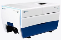
A scalable, high-throughput, high-content screening solution with 7-channel high-intensity laser light source and machine learning capabilities.
-
Titanium dioxide is extensively used in solid dosage forms as an opacifier and colorant. Due to regulatory developments and restrictions by authorities, formulators are increasingly interested in titanium dioxide-free solutions for both dietary supplements and medicinal products. Uniform colored and white coatings applied to tablets not only have an aesthetic function but also improve patient compliance.
-
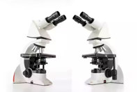
Uniquely Ergonomic System Microscope Leica DM1000
The ergonomically designed Leica DM1000 with adjustable features and easy-to-use controls adapts to every user to improve comfort and convenience while working at the microscope.
Featuring high-quality optical performance, the Leica DM1000 is ideal for all clinical laboratory applications, in particular cytology, haematology, and pathology. For special diagnostics requirements, the microscope is certified for in-vitro-diagnostics (IVD) like in-vitro-fertilization (IVF).
Its advanced capabilities, including fluorescence, make it well suited also for complex tasks.
-
Enhance tablet effectiveness across various dosages with an excipient that offers excellent compressibility and stability for APIs, which eliminates the need for intense processing and provides rapid disintegration.
-
Discover an automated solution that revolutionizes 3D model creation for life scientists to enhance throughput and precision integrated with advanced imaging and analysis systems.


