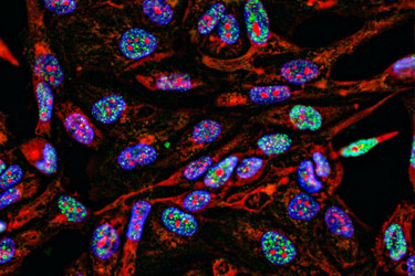
Studying The Spatial Architecture Of Glioblastoma With Highly Multiplex Imaging
By Dr. Rouven Hoefflin

This content is brought to you by Abcam, a Danaher Operating Company.
Cancer cells exhibit significant variability not only between tumors from different patients but also among cancer cells within the same tumor. This latter phenomenon, known as intra-tumor heterogeneity (ITH), influences various aspects of tumor biology, including progression, metastasis, and resistance to therapy. Understanding the causes and consequences of ITH is crucial. Traditionally, pathologists have studied ITH using routine H&E stainings. With the advent of genomics, ITH has also been analyzed through bulk genome and transcriptome sequencing and, more recently, single-cell RNA sequencing (scRNA-Seq). New methods are being developed to add spatial context to scRNA-Seq studies; however, many of these technologies still lack true single-cell resolution.
To bridge this gap, highly multiplex immunofluorescence imaging platforms have been developed to enable the visualization of dozens to potentially hundreds of proteins using conventional antibodies conjugated to unique DNA barcodes (PhenoCycler® Fusion), metals (Multiplexed Ion Beam Imaging), or through bleaching (Cyclic Immunofluorescence). Abcam offers the largest catalog of high-quality carrier-free antibodies (BSA and azide-free), which are crucial for antibody conjugation. This extensive selection allows for the creation of custom antibody panels that target not only canonical epitopes of cells within the tumor microenvironment (TME) but also different cancer cell states that drive ITH.
Access the application note to discover how using a 40-plex antibody panel with the PhenoCycler® Fusion platform allowed investigation into the spatial architecture of glioblastoma (GBM), the most aggressive and common type of brain cancer.
Get unlimited access to:
Enter your credentials below to log in. Not yet a member of Pharmaceutical Online? Subscribe today.
